(Thesis) Analyzing the Softening Behavior of Brain Vessels
University of Utah • Biomechanics • Mechanical Engineering• Experimental Design & Execution
Summary
Cerebral arteries can mechanically "soften" after sub-failure loading, even though they do not show any physical signals of damage. This type of damage is likely occurring during traumatic brain injuries (TBI). However, because the damage is difficult to deteect, sub-failure vessel damage is largely untreated and likely causing the long-term, fatal health concerns that are poorly-understood, but known consequences of TBI.
In this thesis, I further investigated the mechanics underlying sub-failure damage in cerebral blood vessels. I designed, executed, and reported a series of experiments providing evidence that collagen fiber damage occurs at sub-failure stretch levels and contributes to the softening behavior observed in brain vessels under these conditions.
This project page provides a simplified version of my thesis and key takeaways. More detailed information can be found in my published thesis. Follow-up work for this research effort has been completed since my defense, and I am also in the process of submitting this work to the Journal of Biomechanics as a first author (expected release 2026).
Skills Required & Applied
- Protocol & experimental design
- Data collection & management
- Coding (MATLAB, LabView)
- Image processing (motion-tracking, feature detection)
- Quantitative comparisons + statistics
- Biochemical reagent handling
- Data visualization
- Technical writing (thesis + scientific paper)
Background
Why softening in brain vessels matters
Cerebral blood vessels are often overstretched during traumatic brain injuries and surgical procedures. Even without rupture, these events can cause the vessel to "soften". This kind of sub-failure damage is not typically accounted for when treating TBI, despite evidence that vascular damage can occur during TBI may contribute to ischemic stroke risk.
Previous work suggested this softening effect is a result of collagen fibers becoming damaged and "denaturing" at sub-failure vessel stretch limits. However, other work has reported collagen fiber damage to occur at higher strains than where vessel softening begins.
My thesis was aimed to further understand and confirm collagen’s role in vessel softening by directly changing the collagen content of brain vessels and testing how that changes the mechanical outcome. We hypothesized that if collagen is driving sub-yield softening, then partial collagen degradation should measurably shift the softening response even if the collagen is not showing signs of damage/denaturation.


Methods
How I tested collagen's role in softening
I tested our hypothesis following two sets of experiments: (1) I compared overstretch-induced softening in native vs collagen-digested brain vessels, and (2) I continously digested brain vessels of their collagen and measured for corresponding softening effects. The preparation of vessels and processing of the data were distinct between the sets of experiments; however, both sets of experiments followed the same general testing protocol:
- Mount and baseline-test vessels on a custom vessel testing system.
- Apply the sub-failure damage condition via axial overstretch or collagen digestion.
- Re-test baseline response and compute softening from Baseline 1 → Baseline 2.
Step 1
Mount and baseline-test vessels
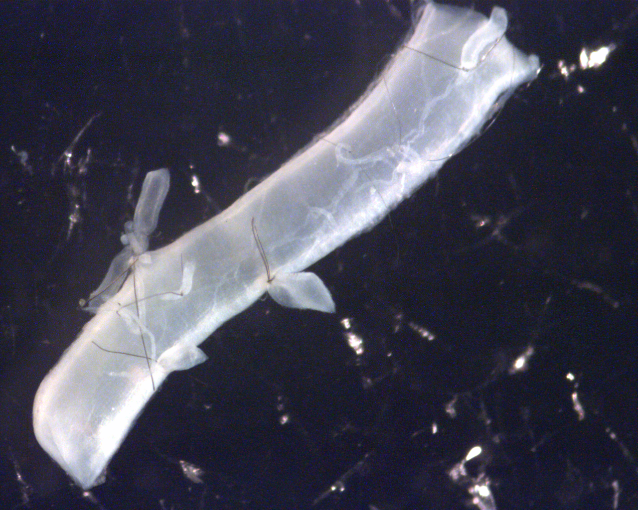
Sheep MCAs were cannulated on hypodermic needles in a PBS bath and secured with sutures and special, benign glue to prevent slip during axial loading. The setup provides axial displacement and force, internal pressure, and synchronized video for deformation tracking.

- (A) Cannulas: Secure the vessel and supply saline fluid to the lumen.
- (B) 3D translation stage: Allows fine axial alignment to ensure the vessel is mounted straight.
- (C) Fluid inlet: Cannula delivering fluid into the vessel.
- (D) Fluid outlet: Carries fluid out of the vessel and can be opened or closed to control pressurization.
- (E) P1 pressure transducer: Measures pressure upstream of the vessel.
- (F) P2 pressure transducer: Measures downstream pressure, allowing pressure differentials to be calculated.
- (G) Actuator guide: Provides slow, precise axial displacement to stretch the vessel, driven by a stepper motor.
- (H) Load cell: Measures axial force with high precision for stress calculations.
- (I) Thermocouple: Monitors bath temperature during collagenase experiments.
- (J) Heating pad: Operates in a feedback loop with the thermocouple to maintain target bath temperature.
Step 2
Apply the sub-failure damage condition
Experiment 1 (overstretch): I extracted cerebral vessels from sheep brains, and digested half of the vessels in collagenase- a biochemical solution that targets and digests collagen fibers. I charactertized a vessel's baseline strength by stretching to its natural length and recorded the force. I then axially overstretched the vessels to 1.1 to 1.4 times the in vivo length and then recharacterized the baseline strength of vessel.
Experiment 2 (continuous digestion): separate cerebral vessels were collected and exposed to collagenase while mechanical response was measured repeatedly over time to characterize how softening occurs with progressive collagen reduction.

Step 3
Evaluation for Softening
Softening was evident in the progressive shallowing of the baseline stress–stretch curves for samples subjected to higher overstretch limits. As expected, collagen-digested vessels exhibited substantially lower stress values across all tests. However, after normalizing the stress–stretch data, both native and digested samples displayed very similar softening behavior.
These results further support collagen’s role in governing vessel softening. The reduced stress magnitudes observed in digested vessels reflect a decrease in load-bearing collagen content. Because digestion was partial, we proposed that the remaining collagen fibers were still dominating the mechanical response, producing softening behavior consistent with that observed in native vessels, as revealed by the normalized data.


Additional Processing
Imaging Damaged Collagen using CHP
Collagen Hybridizing Peptide (CHP) is a molecule that is engineered to bind with damaged collagen fibers. Under a microscopic laser with specific wavelengths, CHP is fluorescent and can be imaged. The intensity of the fluorescence can be correlated with a percentage of damaged collagen fibers.
For this work, we leveraged CHP's abilities, following protocol of other work, to bind CHP to undamaged vessels (control) and our tested vessels (test). The relative increase in intensity of the tested samples were further proof that collagen fibers were damaged.

Results
Main findings
Beyond visualizing the obvious softening occurring between the baseline tests of experiment 1, I quantified softening using common parameterization strategies. Namely, I derived % change in strain energy, in vivo stiffness, dynamic modulus, and in vivo stress after overstretch. The % reduction of these parameters has been shown to correlate strongly with softening in previous work and are standard metrics for quanitifying softening. As expected, the digested vessels showed softening trends practically identical to the native vessels when normalized (see plots in upper right corners). However, the raw softening metrics are much lower in magntitude when compared to the native vessels, which is likely a result of fewer fibers being available to contribute to higher, natural stress values.
In the continuous digestion experiment, mechanical properties degraded progressively over the digestion period, and the resulting “softening-like” patterns were consistent with the idea that collagen governs the observed behavior under sub-yield conditions—even though the mechanism does not require molecular denaturation as the primary explanation.


When these time-dependent digestion trends are compared alongside the overstretch results, the key pattern was found to be consistent: reducing effective collagen contribution shifts the absolute mechanical response downward and reduces the magnitude of softening, while preserving the overall shape of the response under sub-failure conditions.
The comparison figure below summarizes how native vs digested behavior relates across the two experimental approaches, highlighting the interpretation that collagen governs the dominant load-sharing behavior driving softening in these vessels.

Conclusions
Conclusions and future work
This work clarifies collagen’s role in sub-yield overstretch softening of cerebral arteries. Collagen reduction changes the absolute mechanical response and reduces softening magnitude, while the normalized softening pattern versus overstretch remains similar. That combination supports collagen as the dominant contributor without requiring molecular denaturation to be the primary mechanism at the strains where softening begins.
This work is currently being submitted (as of February 2026) for publication to the Journal of Biomechanics.
Sources
References
- M.S. Thesis: Google Scholar citation link
- Lab of Head Injury & Vessel Biomechanics: https://vesselmechanics.mech.utah.edu/research/
- Collagen Hybridizing Peptide (CHP): 3Helix product page
Note: This page is a high-level summary. Full experimental detail and citations are in the thesis/manuscript.